Electrophysiology Study

Overview
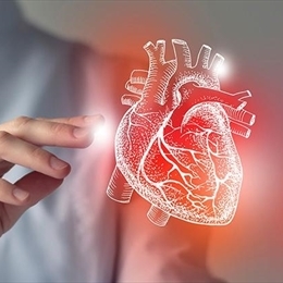
Our heart is an almost entirely muscular organ that has four chambers with heart valves between the chambers. Our heart contracts to pump blood to the body while it relaxes to receive the venous blood to the heart. Contraction and relaxation occur synchronously. On the other hand, the pumping rate or the heart rate is balanced against the oxygen and nutrient requirements of the body. A healthy individual's heart rate is 60 to 100 beats per minute. It beats at a lower limit of this range at resting, while it accelerates above this range in situations that increase the body's oxygen need, such as exercise.
An electrical system within the heart tissue regulates the heart rate and rhythm. Electrical impulses generated by the sinus node and the sinoatrial node are transmitted to the heart through the atrioventricular node, followed by the heart's transmission system (electrical system). Thus, the upper two heart chambers, the atria, contract and pump blood to the lower chambers, the ventricles. Then, the ventricles contract to pump the blood to the body and lungs.
This coordinated pumping function is accurately preserved. The heart rate and rhythm disturbances are observed when a problem occurs in the heart's electrical system. The impaired electrical system may be the usual consequence of aging, but genetic factors and certain medications may also affect the heart rate and rhythm. In addition, the diseases that damage the heart muscle, such as coronary artery disease, also lead to a similar clinical picture.
The electrophysiology study is used to diagnose and treat disorders that are considered to be caused by the heart's electrical system, mainly rhythm problems.
Why is this procedure done?
Failure of the heart to beat within a particular heart rate or in a specific rhythm is manifested by too slow heart rate (bradycardia) or too fast rate (tachycardia), arrhythmia (arrhythmia-atrial fibrillation, atrial flutter) and a combination thereof (bradyarrhythmia or tachyarrhythmia-ventricular or supraventricular).
These disorders cause symptoms such as fainting, chest pain, and palpitations, as well as life-threatening risks such as sudden cardiac death.
In a patient with a documented arrhythmia, location of the electrical system problem is determined by evaluating the heart's electrical system. Up to this stage, EPS is a diagnostic method and the cause of rhythm disorder can be treated with cardiac ablation in the same session.
Risks
Electrophysiology is broadly a safe method, but it does not mean that it poses no risk. The risks that can be faced in an electrophysiology study are listed below:
• Allergic reaction to contrast agent
• Bleeding and/or infection at the catheter insertion site
• Injury of blood vessels
• Damage or perforation of heart
• Injury of heart valves
• Clot formation and migration of the clot to other body parts
• Heart attack
• Stroke
• Death
In the electrophysiology study laboratory, any and all necessary instrument, equipment and other means to manage the possible risks and complications are available.
Our specialists will employ all practices to minimize the risk of complications and our doctors will preoperatively inform you about risks listed above and all other potential complications and will address all your concerns.
Preparation
A cardiologist and/or other healthcare professional will inform you about what you need to do before you visit the catheterization laboratory at the appointment date. The time you will stop eating and drinking will be instructed. Medications you take for diabetes, hypertension and other disorders are reviewed. You are informed about medications that you should take in the day of the procedure. Besides, you should also inform your doctor about all over-the-counter medications, herbal products and vitamin and mineral supplements.
Before the procedure, your health history is reviewed, and a comprehensive physical examination is conducted to evaluate all your vital signs (pulse, heart rate, respiratory rate, body temperature, etc.).
Surgery and early postoperative period
When you are taken to the EPS laboratory for the procedure, you will be placed on the procedure table. Imaging devices are positioned around the table to capture images during the procedure. An IV line is inserted to allow for intravenous treatments and medications as needed. At this stage, a sedative is administered to put you to sleep. All your vital signs are closely monitored using EKG, pulse oximetry, and anesthesia monitoring.
Before the procedure, your health history is reviewed, and a comprehensive physical examination is done to evaluate all your vital signs (pulse, heart rate, breath rate, core temperature, etc.).
Surgery and early postoperative period
After you have been taken to the EPS laboratory for the procedure, you will be positioned on the procedure table. Imaging devices are available around the table to capture images during the procedure.
If necessary, an IV line is inserted to allow intravenous treatments and administering medications. At this stage, a sedative agent is administered to make you fall into a nap.
All your vital signs will be closely monitored using ECG, pulse oxymeter and anesthesia monitor.
Although an artery located in your groin area is more commonly used for EPS, it is possible to use an artery of your arm and your neck.
After the artery that the catheter will be inserted is determined, the local anesthetic is instilled beneath the skin to make sure you do not feel any pain.
Next, a small incision is made over the artery located at the catheter puncture site and a sheath is first inserted. A catheter is inserted into this sheath and advanced to the heart.
A contrast agent that allows vessels to be visualized is administered into the catheter. All heart structures and blood vessels are visualized.
In the EPS study, the heart's electrical activity is first assessed, and measurements are made at various points in the heart. The results of these measurements allow your doctor to map the heart's electrical activity.
Later, your doctor sends some planned stimuli to find the cause leading to dysrhythmia or heart rate abnormality. By repeating this process at various points of the heart, the defect(s) in the electrical and conduction system of the heart is/are determined. After completing this diagnostic phase of the EPS procedure, if your condition allows and your doctor considers it appropriate, the diseased conduction pathways are removed using energy (cardiac ablation). EPS can take about one to six hours, depending on whether a conduction problem has been identified and whether cardiac ablation has been performed.
Next, the catheters are removed, and the procedure is terminated by stitching the small incision.
After completing the EPS, you will be taken to the observation room. Before you are transferred to the patient room, you should be observed here for a while and verify that all your vital signs are stable or within acceptable limits.
Mainly, if the artery located in your groin is used, bleeding should be carefully monitored and managed after the EPS. For this purpose, it may be necessary to apply compression on the small incision made in the groin.
Discharge after an electrophysiology study is entirely related to your health status. You will usually be discharged on the same day if there is no abnormality. But, if an abnormality has been identified and intervened, your cardiologist will want you to stay in the hospital for one night or longer, if necessary.
After discharge from the hospital, if you experience signs of infection such as pain, redness, and swelling at the incision site in the groin, significant swelling or bleeding at the catheter site, and if you experience chest pain or shortness of breath, it is a vital necessity to seek emergency medical treatment immediately.
Results
Your doctor will issue a report after results of the EPS are reviewed.
Depending on the documented problems and whether or not cardiac ablation has been performed, a treatment plan will be prepared.
You should take your medicines, as instructed by your doctor, and comply with health life style recommendations. Do not smoke or quit smoking, if you are a smoker, and you need to maintain optimal body weight and control your blood pressure, blood glucose and blood lipids well.