Oral Diagnosis and Radiology
"Oral Diagnosis" literally means "analysis of the inner mouth." A successful oral treatment is only possible if an accurate diagnosis has been made. Oral diagnosis in dentistry is the practice of determining all problems inside and outside the mouth by using scientific knowledge to determine the relationship between them, thereby helping to make the right decisions regarding treatment based on the findings in hand.
An effective oral diagnosis can be done through oral, external, and radiologic inspections. An extraoral examination includes examining the tissues in and around the mouth (face, jaws, temporomandibular joint, lips, nose, neck, chin…).
Additionally, intraoral examination systematically inspects the teeth. Receding gums, plaque, tartar buildup, abscess presence, mobility, cavities, inharmonious restorations, complications, color, count, and shape defects should all be assessed. Following the inspection of each tooth, the patient should also be examined in terms of closing jaws and their relation with each other. After evaluating all teeth and intraoral tissues, the patient should be informed of any necessary intervention with teeth that don't currently cause discomfort.
Tooth cavities that are not deep may not cause discomfort but can be a source of pain should they progress. Also, being well informed about gum disease in its infancy can prevent receding gums and tooth loss in the future. Oral diagnosis is essential not only in dentistry, but also in terms of systematic diseases. The patient should be inquired regarding frequent diseases. For example, diabetes mellitus, heart disease, hypertension, atherosclerosis, and hormonal disorders, along with the medication used by the patient for these, could affect dental treatment planning. With diabetes mellitus, atherosclerosis, and hypertension, extreme care should be taken during surgical and implant procedures, as the medication used for these diseases directly affects the amount of time it takes for blood to clot. Furthermore, specific intraoral and extraoral findings can be a symptom or indication of certain systematic diseases.
The patient's general health status, health problems relevant to available medical sciences, prescribed medications, and allergic conditions are all factors that may change the dentist's choice of drugs and intervention. Therefore, in the Oral Diagnosis department, an anamnesis containing detailed information about the patient is taken, and treatment is planned accordingly. For example, if the patient has allergic reactions, they may need to be tested before anesthesia. Similarly, the patient may need to consult their dentist and refrain from taking blood thinner medications such as Coumadin, Plavix, or Aspirin for a certain amount before beginning the treatment. In addition to these, a patient's expectations from the treatment should also be assessed. If, for example, the reason for applying to a dentist is to have more aesthetic teeth, the expected result should be thoroughly expressed. By doing this, the doctor can better inform the patient about the applicability of the expectations.
One of the most essential assistive diagnosis techniques in dentistry is radiologic examination. Radiographs used in dental radiology can be divided into intraoral and extraoral.
Inside of the Mouth (Intraoral) Radiographs
Intraoral radiographs encompass periapical radiographs, which many dentists routinely use. With periapical radiographs, one or multiple teeth and their surrounding tissues and the alveolar bone surrounding the teeth can be observed. Tooth decay, dental abscess, periodontal bone loss, secondary cavities, and many more disorders/diseases can be displayed with periapical radiographs.
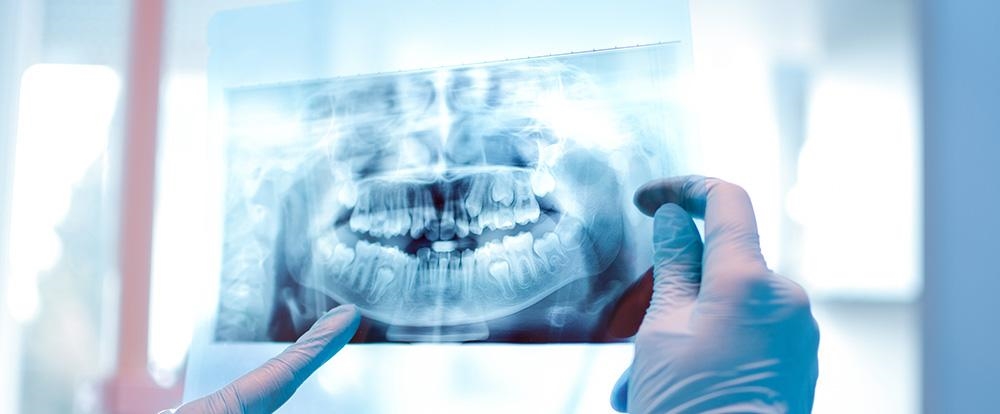
Panoramic Radiographs
Panoramic radiographs are an extraoral imaging technique capable of displaying all teeth, buried teeth, surrounding bone tissue, complete jawbone, physiologic and pathologic gaps in the mouth, and joints at once in a single frame. It is mainly used for imaging simple procedures such as general oral examination, buried tooth extraction, resection, small cysts, or a few implants. While it reflects all teeth simultaneously, it exposes the patient to less radiation than a serial periapical radiograph and allows the doctor to make a general evaluation.
Periapical Radiographs
Allowing doctors to image a small number of neighboring teeth and the bone tissue surrounding them, peripheral radiographs are an intraoral imaging technique used to get a more detailed picture of suspicious circumstances detected in panoramic radiographs. In terms of details, it is much more precise than panoramic radiographs.
Why are panoramic X-rays necessary?
Panoramic x-rays diagnose decays, cysts, and tumors invisible to the human eye. They are radiographs that assist in making treatment planning faster and more accurate. They are images that the doctor must see before maxillofacial surgery, as they display the area in question in detail and increase the success rate of the operation.
What are the advantages of panoramic x-rays?
Panoramic X-rays provide early diagnosis of several cavities as well as cystic and tumor-related formations.
All teeth can be displayed with one single x-ray. An image of all of the teeth allows for early diagnosis and treatment. By this, it is both time and cost-efficient. Standard panoramic radiographs also provide vital information regarding right and left biting radiographs, undetectable midface decay, chronic lesions, buried teeth, problems with the periodontal tissues (bone grafts), formations in the jawbone, excessive fillings, and additionally, in children, the location of permanent teeth, the development of roots and resorption of baby teeth roots. In patients with tooth trauma, immediate and periodic periapical radiographs provide information regarding the tooth, the root, and its surroundings. In patients over 40, regardless of complaints, radiographs are crucial for determining bone pathologies and asymptomatic diseases.
With the radiologic findings, alternative treatment planning can be done more easily once all problems have been determined. In short, a half-hour initial diagnosis will lead to a successful and informative treatment approach.